About the WDP bulb
The very first section of the duodenum, which immediately follows the stomach, is called the bulb. It received such an unusual name because it resembles this vegetable in shape. This department plays an important role in the body. The food we eat is digested in the stomach. To do this, she needs gastric juice, which also contains acid. A lump of food, mixed with gastric juice, enters the duodenum. However, an acidic environment is harmful to the intestines, so in the very first section, the duodenal bulb, the acid is neutralized.
The main function of the onion is to remove acidity, that is, to convert the acidic reaction of food into neutral. Gradually, moving through the duodenum (duodenum), the food mass acquires an alkaline reaction. Food passes through the bulb quite quickly, that is, it takes about 2 or 3 minutes to neutralize it. If food with a too acidic environment enters the bulb from the human stomach, then the bulb cannot cope with its duties, inflammation may appear, and over time, an ulcer or other disease.
Principles of bulbitis prevention
The main preventive measures are a balanced diet, moderation in drinking alcohol, and absolute cessation of smoking. In addition, the stomach and intestines do not tolerate stress and extreme fatigue, therefore, in order to protect them from inflammation, it is necessary to have proper rest and get enough sleep.
Also, timely treatment of pathologies that can provoke a deterioration in the condition of the mucous membrane of the duodenal bulb plays a role in preventing the development of bulbitis. So, it is imperative to treat gastritis and Helicobacter pylori infection before it has time to take root in the initial parts of the intestine. It is advisable to check for worms (if there are any signs of infection) and take special medications if they are detected.
In addition, to prevent the duodenal bulb from being damaged by medications, they must be taken only as prescribed by a doctor. Particular caution should be taken when using nonsteroidal anti-inflammatory drugs, antihypertensive drugs, hormones, antibiotics, and anticoagulants. People take many of these medications every day (for example, aspirin or a pill for high blood pressure), without thinking about the consequences. And they can be very serious.
Zubkova Olga Sergeevna, medical observer, epidemiologist
7, total, today
( 46 votes, average: 4.26 out of 5)
Celiac disease: symptoms and treatment
Travelers' diarrhea: symptoms, treatment, prevention
Related Posts
The main causes of the disease
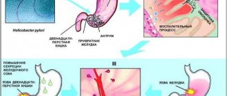
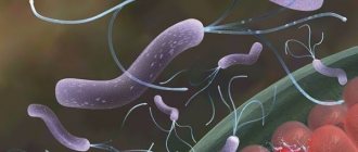
Most often, in 70% of cases, the cause of bulbitis is the bacterium Helicobacter pylori. It is because of this that the catarrhal form of the disease develops. Another fairly common reason is the presence of worms or lamblia. These parasites live in the intestines and affect its functioning; they can cause inflammation of this organ and other diseases. But these are not all the reasons that cause duodenal bulbitis.
If a patient has dyskinesia, that is, a motor disorder of the duodenum, then he also experiences duodenostasis. Partial duodenostasis is bulbostasis, when food mass is retained in the bulb, causing it to increase in size. If this retention of its contents in the intestines lasts long enough, inflammation develops.
Reasons for development
There are quite a large number of reasons due to which bulbitis of the stomach and duodenum progresses, and they are not always pathological. Symptoms of the disease can be triggered by:
- performing surgical interventions on the duodenum;
- long-term use of alcoholic beverages;
- smoking;
- use of medications that have a negative effect on the duodenum;
- poor nutrition and consumption of too spicy foods;
- stressful situations;
- gastritis;
- penetration of infectious agents into the digestive system;
- progression of ulcerative pathologies that have a chronic course;
- weak immune system;
- mechanical, thermal or chemical trauma to the walls of the duodenum.
Causes of stomach bulbitis
Additional reasons
- The patient retained the fetal mesentery. Because of this, the intestine becomes too mobile and forms additional loops. The food mass gets stuck in them, and bacteria begin to multiply rapidly in this mass, which can lead to inflammation.
- The disease develops against the background of gastritis. In some forms of gastritis, large amounts of hydrochloric acid are produced. Some of it ends up in the KDP. This may cause the appearance of superficial bulbitis.
- He developed Crohn's disease. This is inflammation of any part of the gastrointestinal tract or all parts at the same time, which appears for an unknown reason. Then the disease occurs in a chronic form.
- The patient ate a lot of food that irritated the gastrointestinal tract, abused alcohol, and this affected the condition of the duodenum, which is quite sensitive. Because of these reasons, acute bulbitis of the duodenum develops.
- Injuries or taking certain medications can also cause an acute form of the disease. A weakened immune system and constant stress also contribute to inflammation.
- Swallowing objects. If a person accidentally or deliberately swallows any objects, he may develop focal bulbitis. A foreign body enters the bulb and compresses its wall, causing inflammation to appear around this area.
Superficial bulbite
This is inflammation of the duodenal bulb, the mildest form of inflammation. The symptoms are the same as in most diseases of the gastrointestinal tract. Severe stabbing pain in the abdomen and in the navel area. The disease is manifested by vomiting and belching with a bitter aftertaste. Pain occurs an hour or two after eating or with a large time gap between meals. The main treatment is a therapeutic diet for a month, after which you can return to your usual diet.
Varieties
There are several classifications of bulbit. Let's look at the main ones.
Acute or chronic, focal or total
Like many gastrointestinal diseases, depending on the nature of the disease, it can be acute or chronic.
If this is an acute form of the disease, then the symptoms are quite pronounced. Most often the patient has to be hospitalized. In the case when a person does not fully treat acute bulbitis, he develops a chronic form of the disease. He may suffer from it for a long time, several years; periods of exacerbation will be followed by periods of remission. Depending on where exactly the inflammation was localized, a focal form of the disease and a total form of the disease are distinguished. If the patient has focal bulbitis, then a certain area of the bulb becomes inflamed. This disease is easier to cure. If the entire bulb is affected, then this form of the disease is called total. Dealing with it is much more difficult.
Main classification
The depth of damage to the mucous membrane during bulbitis can be different. Depending on it, as well as on the prevalence of the pathology, several forms of the disease are distinguished:
- Atrophic bulbitis. If the patient has not received proper treatment, this form of the disease appears. The mucous membrane gradually becomes gray, its nutrition is impaired, and the duodenal membrane becomes thinner.
- Hyperplastic bulbitis. This also occurs when the disease is advanced. Large folds form and cell proliferation is observed.
- Catarrhal bulbitis. Occurs due to the bacteria Helicobacter pylori. With this form of the disease, the level of acidity changes, which also affects digestion.
- Erosive bulbitis. Small erosions and ulcers appear on the mucous membrane, which can bleed and grow over time. Without timely treatment, it can develop into an ulcer.
- Superficial bulbitis. This is the mildest form of the disease; only the upper part of the mucosa is affected.
- Hemorrhagic bulbitis. Areas of hemorrhage appear on the surface. There may be many of these areas or they may be single.
- Erythematous bulbitis. Red, oval-shaped spots can be seen on the mucous membrane.
- Follicular. Appears due to helminths. Bubbles are visible on the mucous membrane. If left untreated, an ulcer will quickly develop.
Types of bulbite
The disease is classified depending on the course and forms of its manifestation. There are two types - acute and chronic. With timely treatment, the disease is cured completely and without consequences. Neglect of the disease leads to a chronic course. Forms of bulbitis are also distinguished according to the severity of the course, depending on the site of localization of the inflammatory process.
- Moderate gurgling. The pathology is characterized by dull pain and a feeling of heaviness after eating. The pathology causes nausea, vomiting, and frequent belching.
- Superficial bulbitis. The inflammatory process affects only the mucous membrane. As a result of inflammation, pain and swelling appear, and it is difficult for digestive juices to enter the duodenum. The condition leads to stagnation of bile and a lack of enzymes required for digestion. Superficial bulbitis has two forms - acute and chronic. The acute form is infectious in nature, the chronic form is characterized by alternating periods of exacerbation and remission.
- Catarrhal bulbitis. This type of disease is a severe phase that occurs as a result of neglect of superficial bulbitis. Expansion of capillaries on the surface of the mucosa, impaired intestinal motility, reflux, and the release of large amounts of mucus are observed. This type has a seasonal exacerbation. May be asymptomatic. Becomes pronounced as a result of eating spicy foods, stress or drinking alcohol.
- Erosive bulbitis. With this type, deep damage to the bulb tissue occurs. The inflammatory process is observed up to the muscle layer. Erosion is caused by Helicobacter in combination with gastritis. With chronic erosive bulbitis, the patient experiences discomfort after eating, and aching pain appears at night. On palpation, severe pain appears. With erosive-hemorrhagic bulbitis, traces of blood appear in the stool. Neglect of the disease can lead to the formation of ulcers.
- Focal bulbitis is characterized by the spread of the inflammatory process. Foci of inflammation can form both towards the intestines and towards the stomach. This type of disease often manifests itself as a result of hormonal imbalances. Exacerbations are often caused by vitamin deficiency, prolonged fasting, and strict diets.
- Atrophic bulbitis. This species is characterized by the appearance of sour belching, which appears after eating. The patient is worried about heaviness in the abdomen, rumbling, and abnormal stool.
- Follicular bulbitis. This type of bulbitis develops as a result of inflammation of the lymphatic vessels. Small nodular formations – follicles – appear on the inner surface of the duodenal bulb. The causative agents of the infection are helminths and Giardia. Follicular bulbitis responds well to treatment.
Currently reading: What is catarrhal bulbitis of the stomach - types, treatment and proper diet
Symptoms
The symptoms of this disease can be different: sometimes they are barely noticeable, sometimes they are quite bright, it depends on the type of disease.
Symptoms of acute and chronic bulbitis
Often appears after severe poisoning or due to drug abuse. In this case, the symptoms are vivid and it can be difficult not to notice them:
- pain appears, it is quite strong;
- the patient feels sick, he may vomit, sometimes mixed with bile;
- there is a bitter taste in the mouth.
Acute bulbitis can easily be confused with gastroenteritis or pancreatitis in the acute stage.
If it is a chronic bulbitis, then the pain is not so strong, rather aching. Most often they appear due to poor nutrition, for example, if the patient has eaten too much, they can make themselves felt 1-2 hours after eating. Sometimes nausea appears, but without vomiting. The chronic form of the disease lasts several years and has seasonal exacerbations.
Symptoms of different types of bulbitis
Symptoms of the disease largely depend on the form of the disease:
- Erosive bulbitis. The skin is pale, the patient complains of weakness, dizziness, and nausea. Pains that appear at different times of the day are also disturbing; there are also so-called hunger pains.
- Superficial bulbitis. The symptoms are not so pronounced, there may be pain, but it is not very strong. Nausea, but no vomiting, the patient suffers from flatulence, the tongue is covered with a white coating.
- Catarrhal bulbitis. A person complains of poor appetite, weakness, dizziness. He is bothered by pain, heartburn, belching, and may vomit after eating. I also suffer from constipation.
- Follicular bulbitis. The patient has severe pain that often occurs when he is hungry. Worries about nausea and vomiting, belching, diarrhea.
- Atrophic bulbitis. The stomach hurts, heartburn and sour belching appear, the patient either has constipation or diarrhea. A person loses weight quickly.
These are the main signs of this disease. If you experience these or other symptoms, you should contact a gastroenterologist.
Symptoms of bulbitis
The main manifestations of acute bulbitis:
- intense pain in the epigastric region;
- general weakness, drowsiness;
- decreased appetite;
- nausea, vomiting;
- pain on palpation in the stomach area;
- increase in body temperature.
The most common symptoms of chronic inflammation are:
- pain on palpation in the epigastric zone;
- heaviness in the epigastrium after eating;
- belching sour or rotten, possible belching of air;
- tendency to constipation or diarrhea; unstable stools are also possible, manifested by alternating constipation and diarrhea;
- flatulence, spastic pain in the abdominal area;
- weight loss;
- periodically occurring pain (“hungry”, night, early or late, diffuse girdling or clearly localized, in the epigastric region or in the hypochondrium, which depends on the clinical course of bulbitis), relieved by eating, antacid or antisecretory medications;
- additional symptoms in the form of general weakness, sweating, palpitations, shortness of breath, tremor (possible with the neuroendocrine version of bulbitis).
In some cases, bulbitis can be asymptomatic, being detected accidentally during an endoscopic examination for other diseases or at the stage of complications.
Diagnostics
Only a doctor can make an accurate diagnosis. And it will be based not only on the symptoms of the disease, but also on the results of the examination. Without them, treatment cannot be prescribed.
- First, he will palpate the abdomen. If the patient feels tension in the anterior abdominal wall in the epigastric region and pain appears, this may indicate bulbitis.
- X-ray. Helps to consider changes in the duodenum, for example, bulbostasis.
- Duodenoscopy. With its help, it is possible to discern swelling of the mucous membrane, its hyperemia, the presence of erosions, and so on.
- Daily pH-metry. It is necessary to monitor how the process of acid formation occurs during the day.
Other examinations may be prescribed on the recommendation of the attending physician.
Predictions and prevention
If the patient consults a doctor on time, does not self-medicate and follows all recommendations, the prognosis of the disease is favorable. Preventive measures to help prevent bulbitis include:
- dieting on a regular basis;
- cessation of drinking alcohol and smoking;
- stress reduction;
- timely treatment of gastrointestinal diseases (gastritis, etc.);
- Regular visits to preventive examinations with a gastroenterologist.
Treatment
How to treat duodenal bulbitis? The doctor prescribes the treatment. This could be pills or proper nutrition. Diet adherence is mandatory. If this is acute bulbitis, it is better to treat in a hospital. There, if necessary, they will do a gastric lavage to exclude poisoning and prescribe IV drips.
An important part of therapy is giving up bad habits, including smoking. You should avoid stressful situations or take sedatives. This is especially true when treating a chronic form of the disease.
Medications
If a patient has a bulbitis, you cannot do without medication.
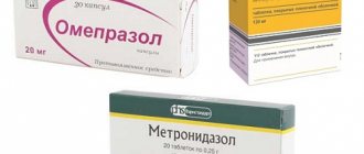
Antacid medications (Gastal, Maalox) will help relieve the attack. But further treatment should be considered by the doctor. So, if this is a catarrhal form of the disease that arose due to Helicobacter pylori, the doctor prescribes antibiotics. He can prescribe enveloping drugs, hydrochloric acid prescription blockers, if it is an erosive form - wound healing agents, and so on.
Diet
In case of acute bulbitis, at first it is better to refuse food, and then you will have to follow a diet. For any form of the disease, part of the treatment is a diet, which should last at least 6 months. After an attack, it is better to eat liquid, warm food. You need to eat up to 6 times a day, in small portions. Slimy porridges, scrambled eggs, chicken broth, compotes, milk, milk soups, and so on are allowed. It is important to limit the amount of salt (up to 5 g per day) and sugar (up to 50 g per day). Bread and other dough products are prohibited.
After 2 weeks, the diet is expanded to include stale bread, butter, cottage cheese, steamed cutlets and pasta. Tea is also allowed, but not strong. All this time you cannot eat salty, smoked, fatty, spicy foods, drink coffee, alcohol, or strong tea.
If treatment was carried out immediately, the prognosis is favorable, but advanced bulbitis is dangerous, as it can cause ulcers, gastrointestinal bleeding, duodenogastric reflux, and so on, so you should definitely visit a doctor.
Treatment with medications
It is quite difficult to cope with duodenal bulbitis without the use of medications. Drugs are prescribed to the patient individually by the attending physician. The specialist builds a treatment regimen based on the results of laboratory and instrumental studies.
The course of treatment for bulbitis usually consists of several areas:
- Anti-helicobacter drugs. Antibacterial agents and proton pump inhibitors (gastroprotectors) are used. As an example, we can cite the following medications: “Sumamed”, “Klacid”, “Flemoxin”, “De-Nol”, “Novobismol”. Antihelminthic therapy. Antiparasitic drugs are prescribed based on the type of helminth pathogen (Praziquantel, Piperazine, Nemozol, Dekaris, Vermox). Antacids. This drug group helps reduce the level of acidity in the stomach and duodenal bulb. Treatment of bulbitis is carried out through the use of drugs “Nolpaza”, “Omez”, “Pariet”, as well as enveloping agents “Almagel”, “Phosphalugel”, “Maalox”. Painkillers and antispasmodics. They are used, as a rule, for severe forms of the disease (“No-Shpa”, “Baralgin”, “Papaverine”). Enzyme preparations. "Creon", "Mezim", "Festal" and their analogues are used as replacement therapy for insufficient production of natural enzymes.
What else to read:
- How to treat acute and chronic gastroduodenitis in adults Contents of the article:1 Causes of the disease1.1 Acute form1.2 Chronic form2 Who is diagnosed with this disease3 Symptoms4 Types of disease5 Diagnosis and treatment5.1 Diet5.2 Medicines Gastroduodenitis is an inflammatory disease,......
- Polyps of the duodenum: symptoms, diagnosis, treatment Contents of the article:1 What is a polyp2 Types3 Causes of polyps in the duodenum4 Symptoms5 Diagnosis5.1 Basic examinations5.2 Additional examinations6 Treatment7 Prevention Duodenal polyp is one of the types......
- Causes and treatment of duodenitis Contents of the article:1 Causes of the disease2 Types of duodenitis3 Symptoms3.1 Acute duodenitis3.2 Chronic duodenitis3.3 Symptoms of different types of duodenitis4 Diagnosis and treatment4.1 Diet4.2 Treatment with drugs4.3 Folk remedies Symptoms are common......
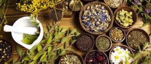
Folk remedies
Good results in the treatment of moderate bulbitis can be achieved with the help of medicinal plants with antimicrobial and anti-inflammatory properties. The use of alternative medicine recipes must be combined with therapy.
St. John's wort decoction
2 tablespoons of St. John's wort herb are poured into 200 ml of boiling water and left for 1 hour. The drink should be taken before meals, 50 g. St. John's wort is prohibited for pregnant women due to the likelihood of miscarriage.
Decoction
You need to take equal quantities of cucumber herbs, cyanosis, fennel, flax seeds, meadowsweet and chamomile and brew 1 tbsp in a teapot scalded with boiling water. l. mixture and 0.5 liters of boiling water. The composition must be infused for 2 hours, then filtered and taken three times a day, 15 minutes before meals, 100 ml. For prevention, it is recommended to carry out a course of 3 months in autumn and spring.
Tincture of rosehip, hawthorn and red rowan
To prepare a medicinal drink, the mixture (1 tablespoon for each ingredient) is poured with 500 ml of boiling water. The composition is infused for 6 hours. Apply 3 times a day before meals, 200 ml.
Infusion of Icelandic moss and chamomile
You need to take 2 tablespoons of plants and pour 0.5 liters of boiling water. The resulting composition should be heated in a water bath for 30 minutes. After time, the liquid is filtered. Use 100 ml three times a day, before meals. Duration of therapy is 2 months.
Currently reading: What is chronic bulbitis of the stomach and duodenum - prognosis and treatment
carrot juice
Only freshly squeezed juice from grated carrots is healthy. You need to take ¼ glass of juice 40 minutes before meals three times a day.
Plantain juice
It is recommended to use juice purchased at a pharmacy. 45 ml of juice should be mixed with 1 tsp. honey and use 1 tbsp. l. three times a day. The duration of treatment is 14 days, after which you need to take a break for 10 days and repeat the course again.
Treatment options
Bulbit, detected in the early stages, does not pose a risk to the patient’s life, therefore, treatment is carried out through conservative therapy on an outpatient basis. In case of exacerbation or bleeding, surgery may be performed.
An example of a daily diet menu for bulbitis
Treatment of duodenal bulbitis is carried out comprehensively. Therapy includes:
- taking medications;
- diet;
- use of folk remedies.
The treatment method is determined only after confirmation of the diagnosis by the attending physician.
Surgical intervention
If the affected areas are bleeding, surgical intervention is performed by esophagogastroduodenoscopy. During the procedure, coagulation or clipping of damaged vessels is performed. Correction of hemodynamic disorders and transfusion of blood components can be carried out jointly.
In the absence of positive dynamics after therapeutic treatment, surgery is prescribed to remove inflamed and necrotic areas of tissue using a polypectomy loop.
In addition to conservative therapy, esophagogastroduodenoscopy may be prescribed
Taking medications
Drug therapy includes taking medications:
- gastroprotectors;
- antispasmodics;
- vitamins;
- antibiotics (if bacteria or viruses are present);
- prokinetics.
Drugs may also be prescribed to prevent and treat diseases resulting from the development of bulbitis.
Diet food
A diet and sample menu for erosive bulbitis are prescribed by a specialist. Patients are recommended diet table No. 1. In case of exacerbation, it is advisable to adhere to nutrition in accordance with menu table No. 0. If the results are positive, the menu is gradually expanded.
To maintain positive results, it is recommended to adhere to proper nutrition for at least six months after the end of treatment. During surgery, the diet is as strict as possible and its duration is long.
Products for the table number 1
Folk remedies
Symptoms of erosive bulbitis of the 12 duodenum and treatment with folk remedies are concepts that are compatible with each other. Traditional medicine methods for treating illness are permissible with the permission of a doctor.
The main direction of this method of therapy involves alleviating the symptoms of the pathology and reducing the intensity of the symptoms that arise.
For medicinal purposes they use:
- infusion of St. John's wort, chamomile or oak bark;
- carrot juice (freshly squeezed);
- flax seed jelly.
Any method of traditional medicine must be discussed with a doctor. Self-medication can cause serious consequences.
The video provides additional information about inflammation of the duodenum:
