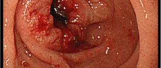
Poorly differentiated adenocarcinoma Adenocarcinoma is a malignant neoplasm that develops from glandular epithelial cells. Poorly differentiated tumors are characterized by a low stage of development of cells, which are primitive cellular elements. Such cells are configured only to consume nutrients and do not perform any functions inherent in healthy tissue. Glandular epithelium is found in various organs, which causes diversity in the localization of adenocarcinoma.
Poorly differentiated adenocarcinoma is a fairly malignant neoplasm that develops from the glandular epithelium of various organs. A low degree of differentiation causes aggressive and rapid growth of the tumor.
Poorly differentiated adenocarcinoma , in which the prognosis is extremely serious, requires timely access to highly qualified medical care. In most cases, the lower the stage of development of adenocarcinoma cells, the more unfavorable the course of the disease.
Prostate adenocarcinoma – what is it?
Prostate adenocarcinoma is a cancer with an increased mortality rate. It occurs as a result of mutation of prostate adenoma cells. At an early stage of onset, the disease is treatable.
Adenocarcinoma mainly affects the peripheral parts of the prostate. Only in 15% of cases does it occur in the central or transitional parts of the organ.
Initially, malignant cells are localized directly in the prostate gland. In the future, they are able to grow into nearby organs. Sometimes the pathological process is limited to the prostate lining. Penetrating into the blood, metastases grow in the bones and lymph nodes of the abdominal cavity.
The main reasons for the appearance
Mutated adenocarcinoma cells differ significantly from healthy ones in structure and function. They do not consume nutrients, however, they multiply at tremendous speed and invade neighboring tissues.
Metastasis begins almost from the very beginning of the formation of a cancer focus and the number of metastases can be significant. It is almost impossible to name reliable reasons why a particular person developed poorly differentiated adenocarcinoma. Experts identify the following predisposing factors:
- abuse of tobacco, alcohol, and narcotic products;
- negative environmental conditions in a person’s place of residence;
- a diet depleted in vitamin composition, with a predominance of preservatives and smoked foods;
- hereditary predisposition – cases of adenocarcinoma already existing in the family;
- working in hazardous conditions;
- prolonged stay in an area with high radiation concentrations;
- abuse of aggressive medications - taking without medical supervision;
- existing chronic infectious pathologies with severe course.
Experts include people in the age category after 55–65 years in the subgroups at risk for the appearance of a malignant lesion; most often, cancer mutations of this form are detected in representatives of the male part of the population. In some cases, it is not possible to establish the true root cause of the malfunction in the body and the further formation of the tumor.
Causes
Adenocarcinoma develops as a result of an uncontrolled increase in the number of atypical cells. To date, the nature of their origin has not been fully studied. But there are a number of factors that increase the risk of developing a pathological process.
These include the following:
- lack of plant foods in the diet;
- prolonged smoking;
- hereditary predisposition to the disease;
- excess weight;
- increased testosterone levels in the body;
- age over 50 years;
- unfavorable environmental conditions.
If there is a hereditary predisposition to malignant tumors, the patient must pay special attention to his health.
Poorly differentiated adenocarcinoma causes
exact reasons for the development of poorly differentiated adenocarcinoma are still not fully understood. With neoplasms of different localization, the following factors may come to the fore in the pathogenesis of the disease:
- benign tumors - malignancy often accompanies the long-term existence of polyps of the stomach and intestines;
- infectious diseases - damage to the gastric mucosa by waste products of the microbe Helicobacter pylori is one of the most important risk factors for the development of malignant neoplasms;
- dietary features - a diet with a predominance of animal fats and meat increases the likelihood of adenocarcinomas of almost any localization;
- hormonal influence - infertility, menstrual irregularities, estrogen replacement therapy, obesity lead to an increase in the concentration of estrogen in the blood and an increase in the likelihood of malignant tumors of the uterus and mammary gland;
- smoking is not only one of the leading risk factors for the development of lung adenocarcinoma, but also increases the likelihood of neoplasms of other localizations, for example, cervical cancer;
- hereditary predisposition - as in the case of other oncological diseases, the tendency to develop poorly differentiated adenocarcinoma, for reasons that are not entirely clear to specialists, can be inherited.
- physical carcinogens - ionizing radiation, magnetic field, vibration, high temperature;
- chemical carcinogens - spicy, salty foods, preservatives, chemical additives, medications;
- biological carcinogens – viral and bacterial infections, parasitic infestations;
- lifestyle factors - unbalanced diet, physical inactivity, gynecological and obstetric history, bad habits.
↑ Top | Applying for treatment ↓
Types of pathology
Prostate adenocarcinoma is classified depending on the degree of aggressiveness. The type of pathology is determined as part of diagnostic procedures. A specific treatment method is selected for each type.
Adenocarcinoma happens:
- acinar;
- moderately differentiated;
- poorly differentiated;
- highly differentiated.
Acinar (small acinar and large acinar)
Acinar adenocarcinoma of the prostate is divided into large acinar and small acinar. In 92% of cases, small acinar adenocarcinoma develops. It is distinguished by a high concentration of mucin content. At the initial stage of its development, multiple small tumors form. Over time, they transform into one large growth.
The division into types is based on changes in prostate tissue, as well as the stage of the lesion and the speed of spread.
Large acinar adenocarcinoma of the prostate is a malignant tumor of the glandular tissue. A diagnosis can only be made through histological examination. In most cases, this type of tumor is fatal.
Moderately differentiated
Moderately differentiated prostate adenocarcinoma responds well to therapeutic manipulations. The process of proliferation of malignant cells is slow. Diagnosis of pathology is carried out during a standard examination by a urologist during palpation. The tumor is localized in the posterior part of the prostate gland.
Poorly differentiated
The most dangerous form of pathology is poorly differentiated adenocarcinoma of the prostate. It is characterized by the rapid growth of malignant cells. This type of disease cannot be eliminated surgically or with the help of medications. The mortality rate is very high.
Highly differentiated
With timely treatment, low-grade prostate adenocarcinoma stops its development. Malignant cells multiply very slowly. This type of tumor can be papillary, mucinous and mucus-forming. Depending on the ability of cells to absorb pigment, adenocarcinoma is divided into dark cell and clear cell.
What does not apply to adenocarcinoma?
Some exocrine gland (exocrine gland) tumors, such as VIPoma or pheochromocytoma, are not usually considered adenocarcinoma. In medicine, such tumors are usually called neuroendocrine.
If the glandular tissue is not malignant or cancerous, the tumor is called an adenoma. The adenoma usually does not invade other tissues and rarely metastasizes. Malignant adenocarcinoma invades other tissues and often metastasizes.
The article was last edited: 04/20/2020
Sources
- Li B., Dai J., Liu S., Gu K., Liu Y., Qu K. (2018). Risk factors and treatment of brain metastases in patients with lung adenocarcinoma. Retrospective analysis based on data from 373 patients. Chinese Neurosurgical Journal.
- Risk factors for breast cancer. Non-profit organization Breastcancer.org, official website.
- Cancer staging (2015). US National Cancer Institute (NCI), official website.
- Risk factors for colorectal cancer (2018). American Cancer Society, official website.
- Signs and symptoms of colorectal cancer (2018). American Cancer Society, official website.
- US R&D Dictionary of Cancer Terms: adenocarcinoma. US National Cancer Institute, official website.
- Risk factors for non-small cell lung cancer (2016). American Cancer Society, official website.
- Signs and symptoms of non-small cell lung cancer (2016). American Cancer Society, official website.
- Survival rates for non-small cell lung cancer (2019). American Cancer Society, official website.
- Risk factors for pancreatic cancer (2019). American Cancer Society, official website.
- Prostate specific gene analysis (2017). US National Cancer Institute, official website.
- Risk factors for brain and spinal cord tumors (2017). American Cancer Society, official website.
Degrees and stages according to the Gleason scale
The Gleason scale is used to classify a tumor according to its degree of neglect. By degree we mean an indicator indicating the intensity of morphological fluctuations in cells. It is assigned after a biopsy. The procedure helps to understand which treatment method will be effective.
The following stages are distinguished according to the Gleason scale:
- First stage. She is assigned up to 5 points on the Gleason scale. There are no symptoms in this case. The pathological neoplasm is localized in one lobe of the gland.
- Second stage. The tumor affects both lobes of the gland. Diagnosis is carried out through ultrasound examination.
- Third stage. Pathological cells grow outside the prostate capsule.
- Fourth stage. It is characterized by metastasis to other organs - the bladder, rectum, sphincter and pelvic walls. According to the Gleason scale, this stage is assigned from 8 to 10 points.
Clinical manifestations of adenocarcinoma
Acinar adenocarcinoma has characteristic clinical manifestations for each stage:
- Stage I - detected very rarely and often suddenly; does not manifest clinical symptoms and is determined by biopsy.
- Stage II - the tumor affects the capsule shell or part of the organ; easily diagnosed using TRUS - the study determines structural changes in the prostate.
- Stage IIIA - adenocarcinoma is actively growing and affects the capsular bag and seminal vesicles.
- Stage IIIB - the tumor has spread to organs adjacent to the prostate.
- Stage IV - adenocarcinoma metastasizes to the pelvic organs and its walls.
For the most accurate and highly informative diagnosis, the determination of the stages of the disease is carried out in conjunction with the Gleason score.
Characteristic symptoms and signs
At the initial stage of development of the disease, there are no specific signs. Until malignant cells begin to multiply intensively, the man has no idea about the disease.
As a result of compression of nearby organs by the nodes, the first symptoms appear. As the tumor grows, they become more intense.
These include:
- feeling of bladder fullness;
- signs of intoxication of the body;
- violation of the ejaculation process;
- a sharp decrease in body weight;
- burning and pain during urination;
- decreased libido;
- increased urge to urinate;
- pain in the perineum.
The course and development of this malignant formation does not differ from other oncological diseases of the male gland in stages.
Clinical picture
Symptoms and signs of the disease completely depend on the anatomical site in which the malignant neoplasm is located.
- When the salivary glands are affected , first of all, pain is noted in the parotid and sublingual area. In the later stages of the process, nodes may be palpated here, usually painful and hard in consistency.
- Adenocarcinoma of the esophagus manifests itself as sharp and severe pain while eating. In addition, patients may complain of a feeling of a foreign body in the chest, as well as a feeling of discomfort in the throat.
- For stomach cancer, the main symptoms are loss of appetite , constant heartburn, difficulty swallowing and pain in the upper abdomen. Often such patients are pale due to a sharp decrease in the level of hemoglobin in the blood.
- Uterine carcinoma may present with watery or bloody vaginal discharge , which worsens during exercise, menstruation, and after sexual intercourse. They often occur when the patient no longer has menstruation.
- Prostate cancer is characterized by severe pain and discomfort in the perineal area, which subsequently leads to impaired urination and decreased potency. If there is a prostate tumor with necrosis, then the urine may change its color.
Diagnostic methods
The effectiveness of treatment depends on the speed of diagnosis. When the first symptoms of the disease appear, you should contact a urologist and describe your complaints in detail.
After collecting information and examining the patient, the doctor will recommend the following procedures:
- biochemical blood test;
- MRI;
- transurethral biopsy;
- ultrasound examination of the abdominal cavity and bladder;
- microslide examination;
- X-ray of the pelvic organs;
- radioisotope research.
Diagnostics
Adequate differential diagnosis of poorly differentiated adenocarcinoma is impossible without laboratory and instrumental examination methods:
Non-invasive procedures:
- ultrasound examination – visualization of dense tissues and cavitary organs;
- X-ray – allows you to identify the pathological focus and its size;
- modern methods of CT, MRI - layer-by-layer examination of organs.
Invasive procedures:
- various blood tests - detailed general, biochemical, and also for tumor markers;
- endoscopic methods – gastroduodenoscopy, irrigoscopy, bronchoscopy, colonoscopy;
- the most informative method is cytological examination of smears and taking biomaterial for biopsy, which allows one to assess the possibility of atypia and its form.
By analyzing all the information obtained from the above diagnostic procedures, the oncologist makes an adequate diagnosis and recommends appropriate treatment procedures.
Treatment
For prostate adenocarcinoma, complex therapy is selected, including medications, surgery and various procedures. The speed of recovery depends on compliance with the rules of therapy. Treatment will be more effective if the disease is diagnosed at an early stage of development.
Hormone therapy
The goal of hormonal treatment is to block the production of androgens, which promote tumor growth. Hormone-based drugs are used in cases where malignant cells grow outside the organ.
If there are metastases, this treatment method will not be effective. In most cases, a man has to take medications for the rest of his life.
The method of treatment is selected by a competent specialist. Self-administration of medications leads to loss of time and aggravation of the situation.
Surgical removal
Surgery is indicated at stages 1 or 2 of the disease. If metastasis has not begun and malignant cells have not had time to affect nearby organs, the prognosis after surgery is favorable.
For prostate adenocarcinoma, I practice the following methods of surgical intervention:
- radical prostatectomy (implies complete removal of the organ);
- excision of the gland without affecting the integrity of the seminal vesicles and capsule.
Sometimes surgery is combined with radiation or chemotherapy.
Radiation therapy
Radiation therapy is used regardless of the stage of the disease. It allows you to reduce the intensity of symptoms, improving the quality of life of a man. In the event that malignant cells are localized only in the prostate area, it is possible to preserve the organ and its functioning.
The following methods of radiation therapy are distinguished:
- Stereotactic. A large dose of radiation is delivered. This allows you to reduce the number of procedures.
- Three-dimensional. The rays affect only malignant cells, without affecting the surrounding area.
- Proton. X-rays are replaced by proton beams.
- With intensity modulation. The technique is considered an improved version of three-dimensional therapy. During the procedure, the device moves around the patient, independently adjusting the dosage of radiation.
Chemotherapy for adenocarcinoma
Chemotherapy is carried out before surgery or in case of ineffective treatment with hormones. It is indicated for metastasis to other organs. To eliminate a tumor in the prostate gland with metastases in the bone or soft tissues, cytostatics or antibacterial drugs are prescribed. They are introduced into the patient’s blood according to an individual scheme.
Aggressive drugs kill not only malignant cells, but also benign ones. Therefore, after treatment, a number of side symptoms occur.
Ablation
The main advantage of ablation is that there is no need to place the patient under general anesthesia. During the procedure, high-frequency current pulses are directed at the tumor. They heat living tissues, promoting the resorption of the tumor.
To avoid damage to healthy tissue, the doctor monitors the process through a monitor. It takes several days for the body to recover after such a procedure.
Cryotherapy for adenocarcinoma
Cryotherapy or cryodestruction is a procedure for freezing malignant cells. It is considered a minimally invasive method of intervention.
As a result of freezing the tumor, its cells stop receiving the necessary nutrition.
The growth of the pathological formation completely stops. After about 2-3 weeks, the body will be freed from dead cells. Cryotherapy can be performed at any stage of adenocarcinoma.
Prevention
No one is immune from the occurrence of cancer. But it is possible to reduce the risk of developing a pathological formation.
Preventive measures include the following:
- It is advisable to adjust your diet. It is recommended to exclude fatty foods, alcohol and fast food.
- It is necessary to periodically drink vitamin and mineral complexes. It is recommended to pay special attention to the content of selenium, zinc and vitamin E in the body.
- Do not neglect preventive examinations by a urologist. Pathological nodes can be detected by palpation.
- Men who lead a sedentary lifestyle are recommended to play sports. This will avoid blood stagnation in the pelvis, thereby reducing the risk of developing pathological neoplasms.
- It is important to treat inflammatory and infectious diseases of the genitourinary system in a timely manner. Because they precede the development of a tumor.
The main differences between adenoma and adenocarcinoma of the prostate
Adenocarcinoma and prostate adenoma are two different diseases that differ in etiology and nature of origin. Adenocarcinoma poses a threat to the patient's life because it is a malignant neoplasm.
Adenoma consists of benign cells. It does not grow beyond the prostate and is not accompanied by metastasis; the symptoms of the diseases are similar.
Adenocarcinoma is treatable, but this requires timely diagnosis. Preventive visits to the doctor help to detect pathology in a timely manner. Therefore, it is extremely important to monitor your sexual health, even in the absence of alarming symptoms.
Related Articles

